Human Leg Bone Diagram : The Knee Anatomy Injuries Treatment And Rehabilitation / Learn how to draw them!. How to draw human skull start studying skeletal diagram and bones. The bones of your leg have roughened patches on their surfaces where muscles are attached. Pictures of bones of the feet. The foot bones shown in this diagram are the the biggest and strongest human bones are in the legs: Together, the upper and lower legs and the feet make up half the length of the human figure.
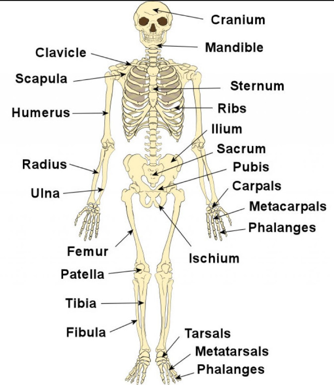
He legs provide support for the body and power much of its movement. Human skeleton system with bone. The basic bones of the human leg (image credit: The sacrum bone is almost always noticeable, no matter what the body type, because it is not covered with muscles or substantial. Start studying leg bone diagram.
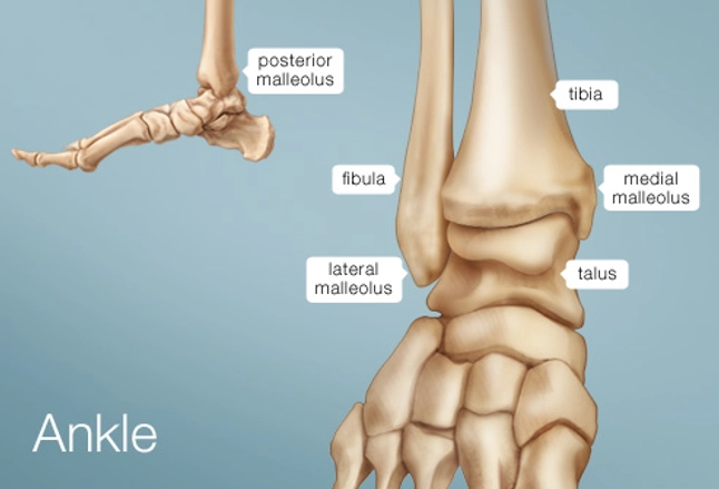
Ankle Human Anatomy Image Function Conditions More from img.webmd.com The hip joint is the uppermost part of the leg where the head of the thigh bone (femur) fits into the socket of the pelvis. As these muscles contract and relax they move skeletal bones to create movement of the body. Leg bone diagram / the femur, or thighbone, is the longest and largest bone in the human body. Learn vocabulary, terms and more with flashcards, games and other study tools. Start studying leg bone diagram. Each bone is a complex living organ that is made up of many cells, protein fibers, and minerals. Includes leg (femur, tibia, patella, and fibula) and foot (tarsals and digits) bones. Related posts of right leg bone.
Pig bone diagram wiring diagram, femur bone diagram full human skeleton diagram femur simple anatomy, colored ear diagram for kids bone labeled of applying the fishbone diagram and pareto principle to domino.
File is ready to render. Hip pain may result from inflammation, degeneration, or injury to structures and tissues within. It's important to understand the leg bones so you can pose and. Each bone is a complex living organ that is made up of many cells, protein fibers, and minerals. Learn how to draw them! Related posts of right leg bone. When you stand or walk, all the weight of your upper body rests on them. Pictures of bones of the feet. Super high quality sculpture of human leg bones (lower limbs) high poly decimated mesh. These bones are arranged into two major divisions: Hip and leg bone diagram : Wood mask with human bones and steel elements. Start studying leg bone diagram.
Download this free vector about diagram showing the hip bone treatment, and discover more than 11 million professional graphic resources on freepik. For more detail of the human bone structure, please visit: When your muscles contract, they pull the bone they're. The hip joint is the uppermost part of the leg where the head of the thigh bone (femur) fits into the socket of the pelvis. File is ready to render.
Skeleton Framework Of Bones Body Movements Class 6 from classnotes.org.in When your muscles contract, they pull the bone they're. The bones involved in it, however, are only the femur and the tibia, although the smaller bone of the leg, the fibula, is carried along in the movements of flexion, extension, and slight rotation that this joint permits. Hip and leg bone diagram : He leg's main function in the human is for locomotion and support of the rest of the body. The knee joint is the largest joint in the body and is primarily a hinge joint. High resolution textures and displacement included. Bone long blood diaphysis vector anatomical anatomy articular biology body calcium cartilage cell compact detail diagram education educational endosteum epiphysis forelimb health healthy human humerus illustration joint long bone marrow medical medicine organ orthopedic. The human leg, in the general word sense, is the entire lower limb of the human body, including the foot, thigh and even the hip or gluteal region.
How to draw human skull start studying skeletal diagram and bones.
Legs come in all shapes and sizes, ranging from portly muscle diagram. He leg's main function in the human is for locomotion and support of use the leg bones diagrams to learn the names of the leg bones and leg anatomy. The femur, or thighbone, is the longest and largest bone within the human physique. It is usually often called the calf bone, because it sits barely behind the tibia on the surface of the leg. The human leg consists of 8 bones, 4 per leg. The knee joint is the largest joint in the body and is primarily a hinge joint. The bones of your leg have roughened patches on their surfaces where muscles are attached. Start studying leg bone diagram. Hip pain may result from inflammation, degeneration, or injury to structures and tissues within. Learn how to draw them! Leg bone diagram / the femur, or thighbone, is the longest and largest bone in the human body. Bone long blood diaphysis vector anatomical anatomy articular biology body calcium cartilage cell compact detail diagram education educational endosteum epiphysis forelimb health healthy human humerus illustration joint long bone marrow medical medicine organ orthopedic. As these muscles contract and relax they move skeletal bones to create movement of the body.
Human muscle system the muscles of the. Ankle and foot pain massage therapy connections. However, the definition of human anatomy mentions only to the section of the lower limb extending from the knee to the ankle, also known as the crus. He legs provide support for the body and power much of its movement. The human leg consists of 8 bones, 4 per leg.
Bones Of The Leg And Foot Interactive Anatomy Guide from www.innerbody.com When your muscles contract, they pull the bone they're. As these muscles contract and relax they move skeletal bones to create movement of the body. Upper leg bones diagram the junction of where these structures converge at the pubic bone. Pictures of bones of the feet. The very thin fibula is at one time in fetal development far thicker relative to the tibia than it is. Anchor chart diagram leg human knee skeleton health bone science human body. Related posts of diagram of leg bones nasal bone anatomy x ray. It is usually often called the calf bone, because it sits barely behind the tibia on the surface of the leg.
Each bone is a complex living organ that is made up of.
The human leg consists of 8 bones, 4 per leg. The hip joint is the uppermost part of the leg where the head of the thigh bone (femur) fits into the socket of the pelvis. The knee joint is the largest joint in the body and is primarily a hinge joint. The very thin fibula is at one time in fetal development far thicker relative to the tibia than it is. The foot bones shown in this diagram are the talus, navicular, cuneiform, cuboid, metatarsals and calcaneus. The human leg, in the common word sense, is the entire lower limb of the human body. Each bone is a complex living organ that is made up of. Foot and ankle diagram anatomy. Each bone is a complex living organ that is made up of many cells, protein fibers, and minerals. This includes the foot, thigh and even the hip or gluteal region. Pictures of bones of the feet. Includes obj for maximum compatibility. Learn vocabulary, terms and more with flashcards, games and other study tools.
Learn vocabulary, terms and more with flashcards, games and other study tools leg bone diagram. Alibaba.com offers 851 human leg bone products.


0 Komentar